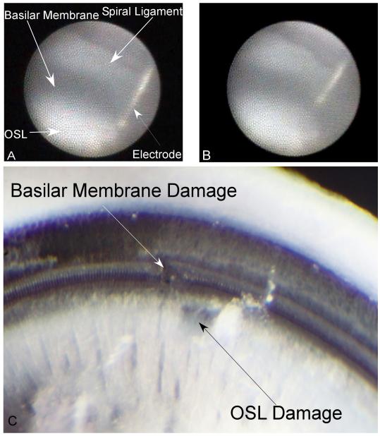
Figure 3.
Gerbil #8, Penetrations 1 and 2: A: Intracochlear structures identified through the microendoscope (right ear) when placed within the round window niche. First penetration. The basilar membrane appears dark due to its transparency to light. The spiral ligament, osseous spiral lamina (OSL) and electrode all appear bright. This image represents the deepest insertion of the electrode in this penetration, where the electrode impacts the OSL. B: Second electrode penetration in the same case. The image is at the deepest penetration depth with the electrode impacting the basilar membrane. C: Corresponding microdissection image indicating distinct damage on the basilar membrane and osseous spiral lamina for the two penetrations.

