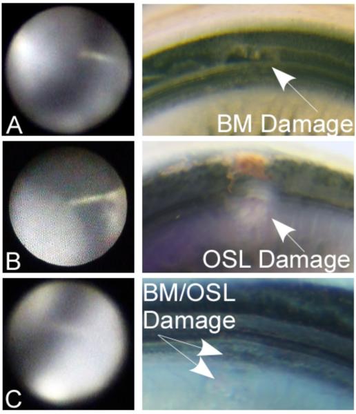
Figure 4.
Representative cases with endoscopic images and correlating microdissections: Case #7; A: Electrode tip directed towards/touching the basilar membrane (BM). The corresponding microdissection demonstrates full thickness damage to the basilar membrane. Case #2; B: Electrode seems directed towards the osseous spiral lamina (OSL). Corresponding histology confirms full thickness injury to the OSL. Case #4; C: Endoscopic image suggesting electrode trauma to the OSL or the junction of the OSL and the BM. Corresponding microdissection shows superficial trauma to the OSL/BM.

