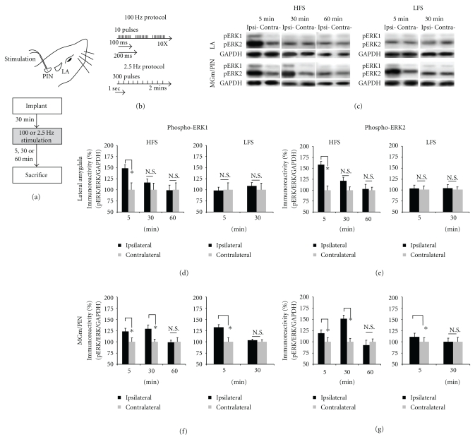
Figure 1.
High-frequency stimulation of the MGm/PIN promotes ERK phosphorylation in LA at 5 min and in the MGm/PIN at 30 min after stimulation. (a) Placement of stimulation electrode and schematic representation of the experimental protocol. (b) Schematic representation of the HFS and LFS stimulation protocols. Anesthetized rats were given HFS or LFS and sacrificed at 5 min, 30 min or 60 min after stimulation. (c) Images of Western blots for phospho-ERK1/2 and associated GAPDH loading controls from LA (upper) and MGm/PIN (lower) samples after HFS or LFS. (d-e) Mean (±SEM) percent phospho-ERK1/2 immunoreactivity from LA punches taken from rats receiving HFS (left) or LFS (right) and sacrificed at 5 min (HFS: n = 6; LFS: n = 6), 30 min (HFS: n = 6; LFS: n = 8), or 60 min (n = 6). (f-g) Mean (±SEM) percent phospho-ERK1/2 immunoreactivity from MGm/PIN punches taken from rats receiving HFS (left) or LFS (right) and sacrificed at 5 min (HFS: n = 6; LFS: n = 6), 30 min (HFS: n = 5; LFS: n = 5), or 60 min (n = 6). For each figure, phospho-ERK1/2 levels have been normalized to total-ERK1/2 levels for each sample and counts on the ipsilateral (stimulated) side have been expressed as a percentage of those on the contralateral (nonstimulated) side. *P < .05 relative to the ipsilateral side N.S. = not significant.

