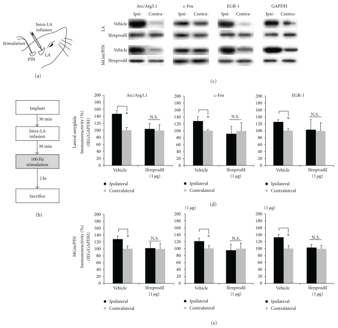
Figure 6.
Pharmacological blockade of NMDAR-driven synaptic plasticity impairs ERK-driven IEG expression in both the LA and MGm/PIN following HFS. (a) Placement of stimulation electrode and infusion cannula. (b) Schematic representation of experimental protocol. Rats were given intra-LA infusion of the vehicle or 1 μg ifenprodil followed 30 min later by HFS of the MGm/PIN. Rats were sacrificed 2 hours after stimulation. (c) Images of Western blots for Arc/Arg3.1, c-Fos, EGR-1 and GAPDH from both LA (top) and MGm/PIN (bottom) samples. (d) Mean (±SEM) percent Arc/Arg3.1, c-Fos and EGR-1 immunoreactivity from LA punches taken from rats given intra-LA infusion of 2% HBC-saline (vehicle; n = 8) or 1 μg/side ifenprodil (n = 8). (e) Mean (±SEM) percent Arc/Arg3.1, c-Fos and EGR-1 immunoreactivity from MGm/PIN punches taken from rats given intra-LA infusion of 2% HBC-saline (vehicle; n = 8) or 1 μg/side ifenprodil (n = 8). In each figure, IEG levels have been normalized to GAPDH for each sample, and IEG expression on the ipsilateral side has been expressed as a percentage of that on the contralateral side for each rat. *P < .05 relative to the ipsilateral side N.S. = not significant.

