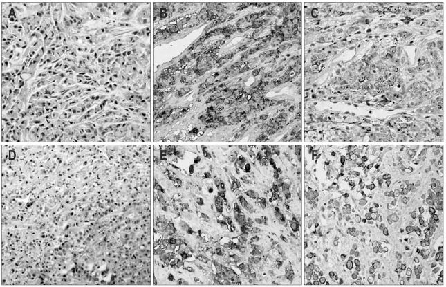
Fig. 3.
(A) Histopathologic findings of the skin biopsy sample showing signet-ring-cell carcinoma infiltration (H&E stain, ×200). (B) The tumor cells in skin mass were positive for carcinoembryonic antigen (CEA) (×200). (C) The tumor cells in the skin mass were weakly positive for CK 19 (×200). (D) Histopathologic findings of the liver biopsy showed poorly differentiated adenocarcinoma infiltration (H&E stain, ×100). (E) The tumor cells in liver mass were positive for CEA (×200). (F) The tumor cells in liver mass were weakly positive for CK 19 (×200).

