
Figure 2. CPG2 Is Specifically Localized to the Postsynaptic Side of Excitatory Synapses.
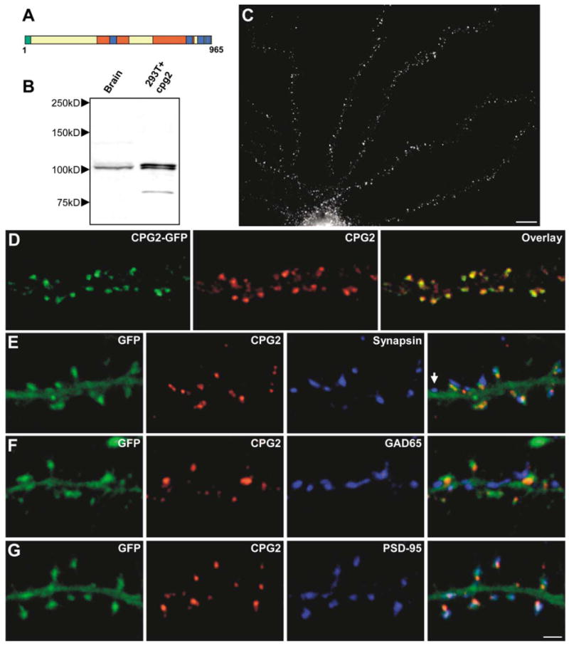
(A) Schematic of the CPG2 protein. Green, additional amino acids in CPG2B; red, spectrin repeats; blue, coiled coils.
(B) Western blot of protein extracts from rat cerebral cortex (lane 1) or 293T cells expressing cpg2 (lane 2) probed with anti-CPG2 monoclonal antibody 200A6.
(C) Cultured hippocampal neuron (24 DIV) labeled with the anti-CPG2 monoclonal antibody. Scale bar, 10 μm.
(D) Cultured hippocampal neuron infected with a CPG2-GFP-expressing lentivirus labeled for CPG2.
(E–G) Neurons infected with a GFP-expressing lentivirus were double labeled for CPG2 and synapsin I (E), GAD65 (F), or PSD-95 (G). Arrow in (E) indicates synapse without CPG2 labeling. Scale bar, 2 μm.