
Figure 3. CPG2 Localizes to a Subdomain of Excitatory Synapses.
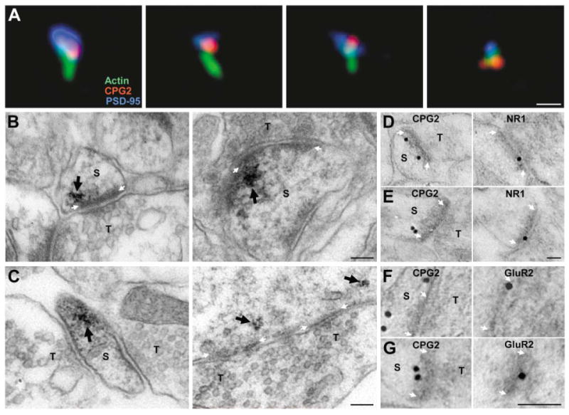
(A) Individual spines from cultured neurons stained for actin filaments (green), CPG2 (red), and PSD-95 (blue). Scale bar, 1 μm.
(B and C) Preembedding CPG2-labeled immunoEM micrographs of synapses from rat dentate gyrus or cultured hippocampal neurons. CPG2 labeling (arrow) is lateral to and underneath the PSD at synapses in vivo (B) and in vitro (C). Scale bars, 100 nm.
(D–G) Postembedding CPG2 and NR1 (D and E) or GluR2 (F and G) immunogold EM on serial sections of rat dentate gyrus. Scale bars for all images, 100 nm. S, spine; T, presynaptic terminal. White arrows mark the PSD.