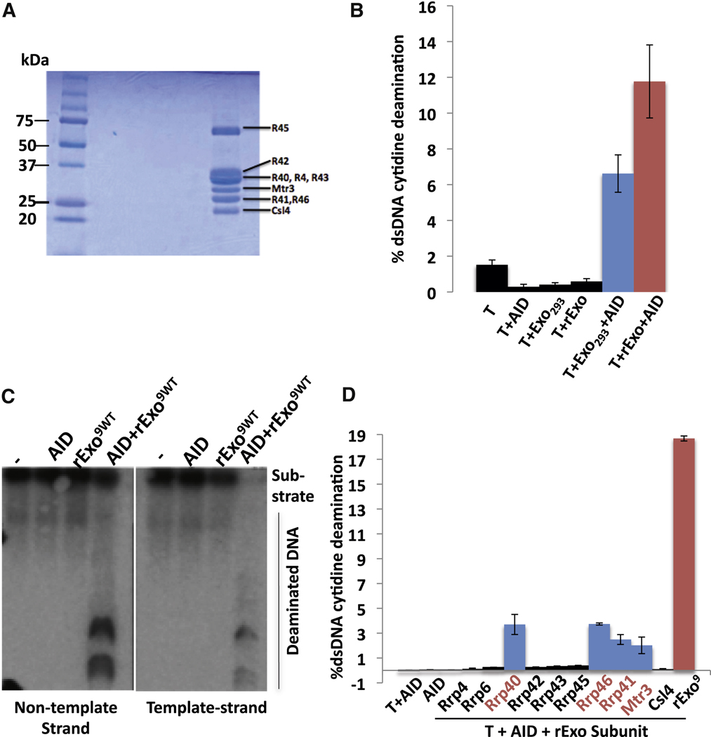
Figure 6. Recombinant core RNA exosome and individual subunits stimulate AID activity.
(A) Coomassie blue stained polyacrylamide gel electrophoreisis analysis of the 9 subunit recombinant RNA exosome complex generated from individual subunits. (B) Comparative analysis of the ability of RNA exosome complex from 293T cells (Exo293) and recombinant RNA core exosome complex (rExo) to stimulate AID deamination activity as measured by 3H release/SHM substrate assay in Fig. 5A. See Supp. Methods and Fig. S6 for details. Optimal amounts of RNA exosome293T and recombinant RNA exosome core complex were used (Fig. S6; Supp. Methods). (C) Assay of recombinant exosome complex to stimulate strand-specific AID deamination of a transcribed SHM substrate via assay in Fig. 5C. Non-template and template strand deamination are shown on left and right panels, respectively. Reactions contained either AID, recombinant core RNA exomsome (rExo9wt) or both (AID + rExo9wt) as indicated. (D) Individual recombinant RNA exosome subunits were assayed for ability to promote AID deamination of a T7-transcribed SHM substrate as outlined in Fig. 5A. Added exosome components are indicated. Control reactions with AID alone or AID plus T7 polymerase are on the left. A positive control with complete recombinant core RNA exosome (rExo9) plus T7 and AID is on the right. For panels B and D, values represent the average and standard deviation from the mean for three independent experiments. See also Figure S 6&7.

