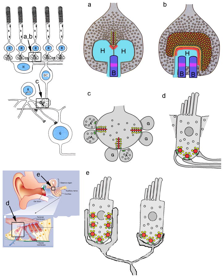
Figure 1. Diversity of ribbon morphology and postsynaptic architecture in different cell types.
a, b: Schematic diagram of transverse (a) and en-face (b) views of a mammalian photoreceptor ribbon synapse117, whose context in the retina is illustrated in the diagram to the left. c: Synaptic arrangement at bipolar cell ribbons. The location of this synapse in retinal circuitry is illustrated in the diagram to the left. d: Synaptic arrangement at ribbons of cochlear inner hair cells. e: Vestibular afferents receive synaptic inputs from multiple ribbons of multiple hair cells. The locations of cochlear inner hair cells and the vestibular apparatus are shown schematically in the diagram to the left. Dark red areas, synaptic ribbons; bright red areas, AMPA receptors; orange areas, arciform density; yellow circles, vesicles attached to ribbon; green circles, docked vesicles; light blue areas, horizontal cell dendrites (H); dark blue areas, ON bipolar cell dendrites (B); magenta areas, mGluR6 receptors; G: ganglion cell dendrite; A: amacrine cell process.

