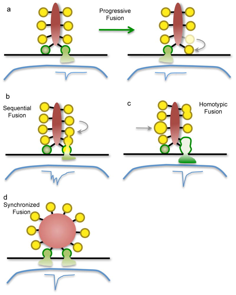
Figure 2. Modes of synaptic vesicle fusion at ribbon synapses.
Green vesicles represent vesicles that are docked at the plasma membrane (black line) at release sites. Blue traces represent the postsynaptic current. a: Progressive fusion, in which primed vesicles docked at different release sites fuse progressively during maintained depolarization, each producing an independent unitary postsynaptic current. Vesicles in higher rows move along the ribbon to replenish empty release sites. b,c: Two forms of compound fusion. In sequential fusion (b), vesicles fuse in sequence up the face of the ribbon, starting with the vesicles docked at the plasma membrane. This produces a burst of quantal postsynaptic currents in rapid sequence. Vesicles attached to the ribbon might fuse with each other (homotypic fusion; c) prior to undergoing exocytosis. The resulting larger bolus of glutamate produces a larger postsynaptic current. d: Synchronized fusion of multiple docked vesicles, which results in a multiquantal postsynaptic current.

