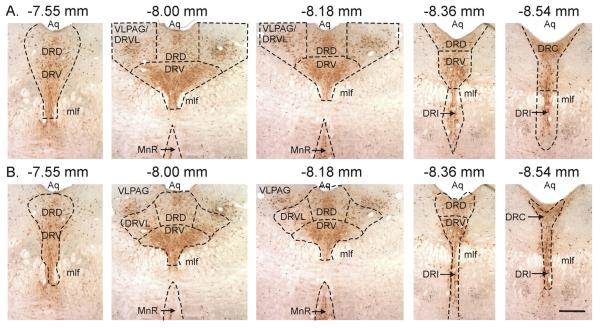
Figure 1.
Topography of midbrain raphe nuclei studied. A) Areas where cell counts were made compared to B) subregions of the DR according to a standard stereotaxic atlas of the rat brain (Paxinos and Watson, 1997). Note in B) how there are substantial numbers of serotonergic neurons which are in the VLPAG rather than in the DR. Scale bar, 400 μm. Abbreviations: Aq, cerebral aqueduct; DRC, dorsal raphe nucleus, caudal part; DRD, dorsal raphe nucleus, dorsal part; DRI, dorsal raphe nucleus, interfascicular part; DRV, dorsal raphe nucleus, ventral part; DRVL, dorsal raphe nucleus, ventrolateral part; mlf, medial longitudinal fasciculus; MnR, median raphe nucleus; VLPAG, ventrolateral periaqueductal gray.

