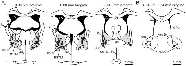
Figure 1. Histological verification of cannulae placement represented on coronal brain section illustrations.
Representative cannulae placements throughout A) a rostra-caudal extent of the BNST (Panel A) and nucleus accumbens control injections (Panel B). The gray areas in Panel A represent the BNST area. Each black dot represents an injection site from each experiment which resides within the BNST area. Black dots in Fig. 1B represent control injection sites in the nucleus accumbens. The distance from Bregma is indicated above each coronal brain section illustration. A 1 mm scalebar is located at the bottom right of Panel A and B. The coronal demarcations are adapted from a standard rat brain stereotaxic atlas (Paxinos and Watson, 1986). Abbreviations: 3V, 3rd ventricle; ac, anterior commissure; aca, anterior part of anterior commissure; AcbC, nucleus accumbens core; AcbSh, nucleus accumbens shell; BSTL, bed nucleus of the stria terminalis, lateral division; BSTM, bed nucleus of the stria terminalis, medial division; cc, corpus collosum; CPu, caudate putamen; f, fornix; ic, internal capsule; LV, lateral ventricle; ox, optic chiasm; sm, stria medullaris of the thalamus; st, stria terminalis.

