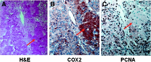
Figure 2.
COX2 and PCNA immunoproteins are observed in both chief cells and oxyphil cells of hyperplastic PTGs from hemodialysis patients. Immunohistochemistry and H&E staining in consecutive sections of the PTG show (A) H&E staining to differentiate chief cells and oxyphil cells (eosinophilic cells, red arrow), (B) cytoplasmic positivity for COX2 in oxyphil cells and chief cells, and (C) nuclear positivity for PCNA in oxyphil cells and chief cells. Magnifications: ×400.

