Figure 3.
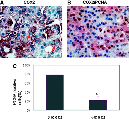
Most COX2-positive PTG cells are proliferative cells. (A) The immunohistochemistry detected some COX2-positive cells in the hyperplasia parathyroids with two nuclei (red arrow). (B) COX2 (red) and PCNA (brown) co-staining in the paraffin-embedded sections of hyperplastic PTGs from hemodialysis patients. Magnifications: ×400. (C) Percentage of PCNA-positive cells in COX2-positive or COX2-negative PTG cells per microscope field. cP < 0.001 versus COX2-positive cells.

