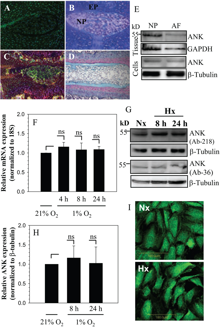
Figure 1.
A–D. Sagittal sections of the intervertebral disc of embryonic (A and B) and a mature (C and D) animal. Sections were treated with ANK antibody (A and C), or counterstained with hemotoxylin, eosin and alcian blue (B and D). ANK was expressed in nucleus pulposus (NP) of the adult and the neonate (see arrow). Annulus fibrosus (AF) and endplate cartilage (EP) (Mag. X 20). E. Western blot of ANK expression in rat disc tissues and cultured cells. F. Real-time RT-PCR analysis of ANK expression by NP cells in hypoxia (1% O2). There was no significant change in ANK mRNA expression in hypoxia. G. Western blot analysis of ANK expression in NP cells in hypoxia using two ANK antibodies. A prominent ANK band (54.4 kD) was detected. H. Densitometric analysis of ANK expression on multiple Western blots. Note, there was no significant change in ANK expression in hypoxia. I, Immunofluorescent analysis of NP cells cultured in hypoxia. There was a similar level of ANK expression in normoxia and hypoxia, Mag X 20. Values shown are mean ± SE from three independent experiments, * p < 0.05, ns = non significant.

