
Fig. 2. System resolution and multiplexed cell culture imaging.
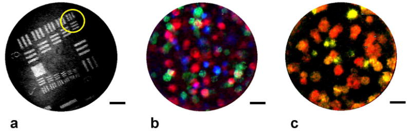
(a) Image of a standard US Air Force resolution target, demonstrating resolution of the 4.4 μm-wide bars in group 6, element 6 (circled). (b) False-color composite image of a 3-D collagen construct containing 1483 oral squamous cell carcinoma cells (red), SK-BR-3 breast cancer cells (green), and SiHa cervical cancer cells (blue), with each cell type stained with a spectrally-distinct fluorophore. (c) Single frame image of the same tissue construct acquired in real-time with a single long-pass emission filter. 1483 cells appear red, SK-BR-3 cells yellow, and SiHa cells appear light green. All scale bars represent 50 μm.