
Figure 2.
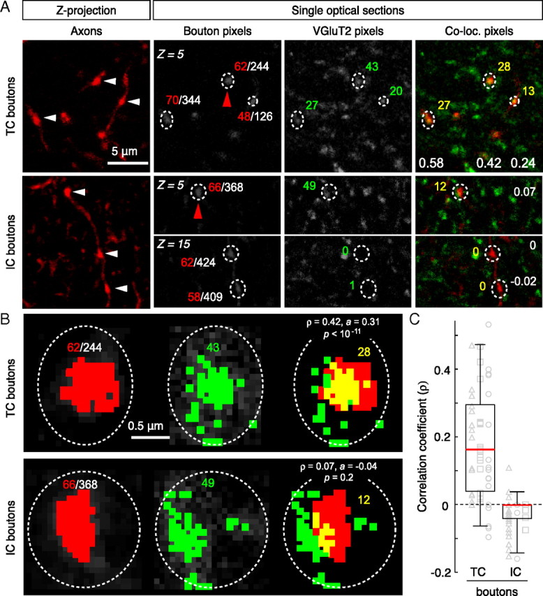
Colocalization analysis in layer 4. A, Examples of micro-ruby-filled TC axonal boutons (arrowheads) in maximum-intensity Z-projections of 15 optical sections (Z = plane number). The grayscale images show single optical sections for each channel and regions of interest with corresponding pixel areas (white denominator: total pixels within region; colored numbers: red, axon; green, VGluT2; yellow, overlap). The white numbers in the colocalized panels are the respective ρ-bouton values. B, Higher magnification views of single boutons from the above panels (indicated by red arrowheads in A). The colored pixels have gray values above the threshold (see Materials and Methods). Note that there is a significant spatial correlation between red and green pixels for the TC bouton, but not the IC bouton. Although ρ is positive for both examples, colocalization is only significant for the TC bouton and is an example of how spurious overlap can be discerned with a relatively straightforward pixel-based analysis. C, Summary of colocalization analysis for each bouton population. The box plots show the distribution of correlation coefficients calculated for each bouton class. The horizontal lines mark the 25th, 50th (red), and 75th percentiles, and the ends of the whiskers mark the 2.5th and 97.5th percentiles (n = 47 TC, n = 50 IC; n = 3 animals per group). The two distributions were statistically different from each other (p < 10−13, K-S test). Correlation coefficients for individual boutons are plotted as gray symbols; data from each animal are represented by different shapes.