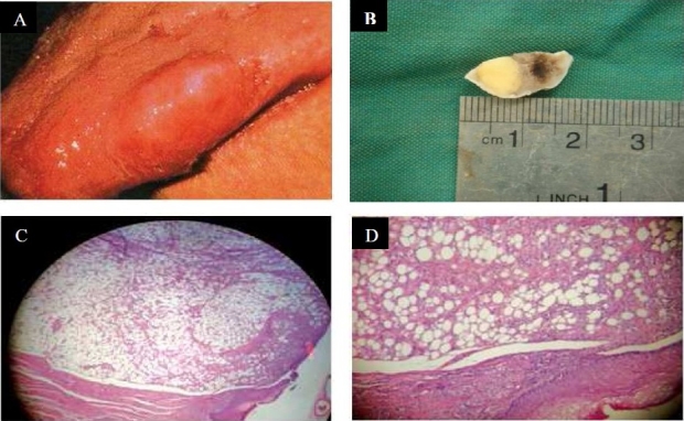
Figure 2.

(A) Submucosal mass on the lateral border of the dorsal part of the tongue. (B) Gross picture of lipoma on the dorsal part of the tongue. (C) Lobulated mass with fibrosis capsule (H & E staining, original magnification X100). (D) Mature fat cells (H & E staining, original magnification X400)
