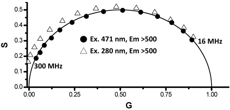
Figure 6.
Decay data of EGFP excited at 471 nm (circles) and 280 nm (triangles). All of the data recorded with 280 nm excitation fall outside the universal circle, which is an indication of an excited state process, allowing for a simple visualization of the energy transfer process between the lone tryptophan (donor) and the EGFP chromophore (acceptor).

