Fig. 1.
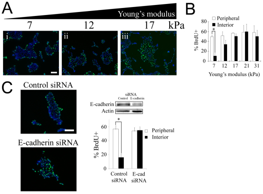
Substratum compliance affects spatial patterns in cell-cycle activity and contact inhibition of proliferation. (A) BrdU incorporation (green) and DAPI staining (blue) were assessed in serum-starved MDCK cells seeded on collagen-coated gels of varying stiffness following 16 hours of treatment with 100 ng/ml EGF. (B) The graph shows the percentage of peripheral and interior cells undergoing DNA synthesis. Values are mean ± s.d. (n=2–5). *P<0.01 (Student's t-test) for comparison of the percentage of BrdU incorporation among peripheral versus interior cells. The lines connecting bars denote the pair of data points being compared in the Student's t-test. (C) Downregulation of E-cadherin eliminates the spatial pattern in proliferation on soft substrates. MDCK cells grown on soft substrates (7 kPa) were transfected with control siRNA or siRNA against E-cadherin and the percentage of peripheral and interior cells incorporating BrdU was quantified following EGF stimulation as in A. The extent of E-cadherin knockdown was determined by western blot. Equal loading was confirmed by probing for actin. Values are mean ± s.d. (n=2). *P<0.01 (Student's t-test). Scale bars: 100 μm.

