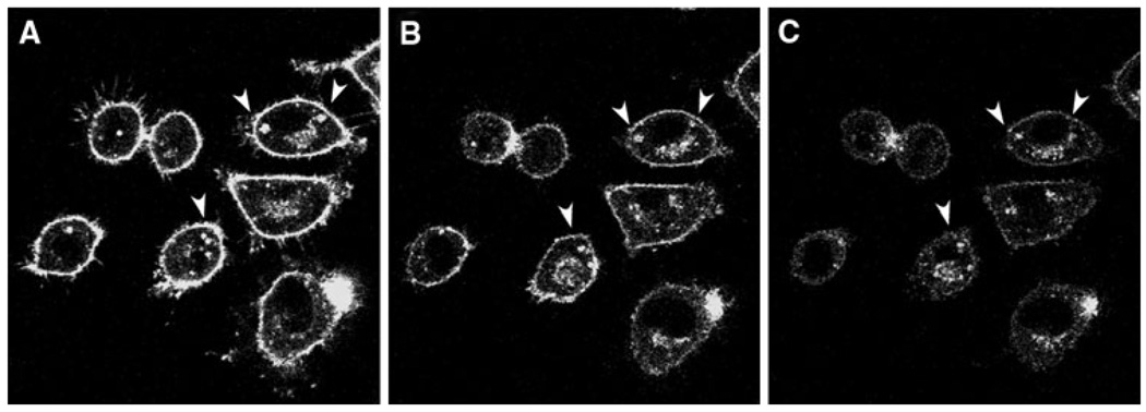
Fig. 2.
HDL-mediated NBD-sphingomyelin efflux in L-cell fibroblasts. Cells were labeled with NBD-sphingomyelin (0.2 µM) and incubated with HDL (10 µg HDL/mL) to begin the efflux process. a 0 min, b 20 min, and c 30 min after the addition of HDL. The arrows indicate high-retention lipid droplets during the time course. Cells were examined with the Bio-Rad MRC-1024 confocal imaging system. Objective, 63×

