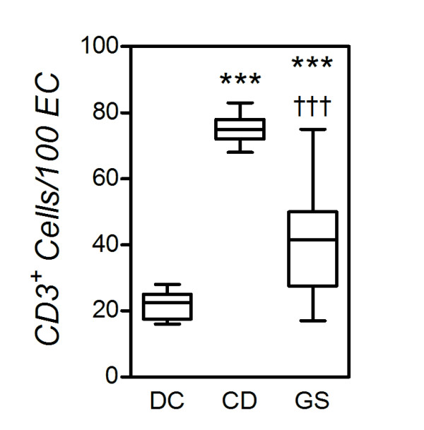
Figure 3.

Reduced numbers of intraepithelial lymphocytes (IELs) in gluten-sensitive patients (GS) versus celiac disease patients (CD). Small intestinal bioptic specimens were stained for the T-cell marker CD3 in immunohistochemistry, and CD3+ cells are enumerated as indicated in Methods. Boxes represent the median (interquartile range), and whiskers represent the range of IEL numbers relative to 100 enterocytes in the same samples in 16 GS, 11 CD and 12 dyspeptic controls (DC). ***P < 0.0005 relative to DC; †††P < 0.0005 relative to CD (Mann-Whitney U test).
