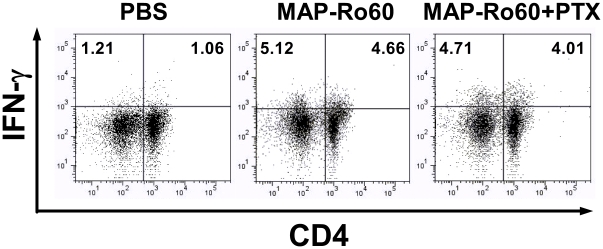
Figure 5. Increased IFN-γ releasing lymphocytes in salivary gland associated lymph nodes from SJL/J mice.
The SG lymph nodes cells isolated from mice immunized with PBS (mock) or MAP-Ro60 +/- PTX were analyzed by flow cytometry assay for IFN-γ+ and specifically CD4+ IFN-γ+ (Th1) lymphocyte induction 10 days post primary immunization. The SG lymph node cells from one group (N = 10) are pooled and divided into 2 samples. Data shown is from one representative experiment.

