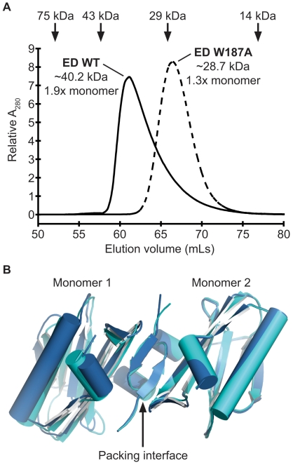
Figure 3. Structural analysis of PR8 NS1 ED W187A.
(A) PR8 NS1 ED-W187A is monomeric in solution. Gel filtration analysis of the purified WT and W187A mutant 6His-tagged PR8 NS1 ED proteins at 1mg/mL. The elution volumes of protein standards used to calibrate the column are indicated at the top: conalbumin (75000 Da), ovalbumin (43000 Da), carbonic anhydrase (29000 Da) and ribonuclease A (13700 Da). The estimated MWs of WT and W187A PR8 EDs are shown together with their ratio to the calculated MW of the respective monomeric protein. Calibrations and calculations were performed as previously described [28]. (B) Crystals of PR8 NS1 ED-W187A form by strand-strand packing. Two crystal forms of the PR8 NS1 ED W187A monomeric mutant revealing strand-strand packing in the crystal lattice. The two monomers of each form are colored dark or light blue (PDB IDs 3O9Q and 3O9R, respectively).

