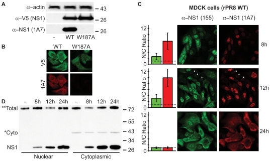
Figure 6. Possible visualization of ‘helix-open’ and ‘helix-closed’ NS1 ED conformational states in virus-infected cells.
(A) mAb 1A7 does not recognize NS1-W187A. Western blot analysis of lysates from 293T cells transfected with pCAGGS expression plasmids encoding the indicated V5-tagged PR8 NS1 protein (or vector only). NS1 was detected using both rabbit anti-V5 pAb and mouse mAb 1A7. Actin served as a loading control. Molecular weight markers (kDa) are indicated to the right. (B) Indirect immunofluorescence analysis of NS1 proteins in MDCK cells transfected with pCAGGS expression plasmids encoding the indicated V5-tagged PR8 NS1 protein. Cells were fixed approximately 24 h post-transfection. Co-staining was performed using rabbit anti-V5 pAb and mouse mAb 1A7. (C) mAb 1A7 and pAb 155 highlight different NS1 populations during infection. Indirect immunofluorescence analysis of NS1 protein localization in MDCK cells infected for the indicated times with rPR8 WT virus (MOI of 2 PFU/cell). Co-staining was performed using rabbit anti-NS1 pAb 155 and mouse mAb 1A7. Asterisks indicate example uninfected cells to show background staining. Bar graphs represent the mean nuclear/cytoplasmic (N/C) ratios of mean fluorescent intensities derived from manually assigned individual nuclei and cytoplasms for each fluorescent channel (n = 25 cells per timepoint, error bars represent standard deviations). The dotted line indicates a nuclear/cytoplasmic ratio of 1. (D) Western blot analysis of nucleo-cytoplasmic extracts prepared from MDCK cells infected as for (C). The primary antibody was a polyclonal rabbit anti-serum raised against a GST-NS1 (RBD) fusion protein. *Cyto indicates a non-specific band that co-purifies solely with the cytoplasmic fractions and thus highlights purification integrity. **Total indicates a non-specific band found in all fractions that serves as a convenient loading control. Molecular weight markers (kDa) are indicated to the right.

