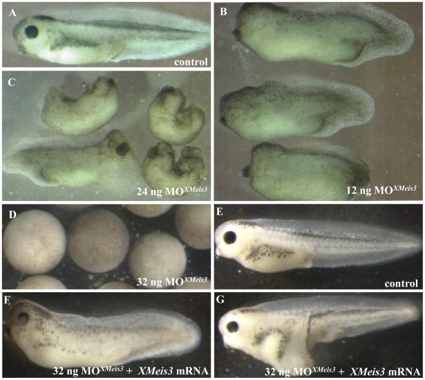
Figure 3. Effects of XMeis3 MO loss-of-function on embryonic development and the rescue of MOXMeis3.
Embryos at the one-cell stage were injected into the animal hemisphere with MOXMeis3 in amounts of 12 ng (B), 24 ng (C), and 36 ng (D), and allowed to develop until the control embryos (A) reached tadpole stages. This treatment disturbs development of the embryonic axis. At the highest concentration, the embryo is blocked during gastrulation (fig. 3D) and then disintegrates to a mass of dissociated cells contained within the vitelline membrane (not shown). The specificity of MOXMeis3 is shown by the rescue with XMeis3 synthetic mRNA. Embryos were injected with 32 ng of MOXMeis3 and 125 pg synthetic mRNA for XMeis3 and allowed to develop until the control embryos reached the tad pole stage (E), In the majority of the embryos a large part of the axis was rescued (F), in a small number of embryos the phenotype could even be reversed, not only is the axis fully rescued but the embryo shown in (G) even possesses additional trunk structures as was revealed by the presence of somites in the axis outgrowth (not shown). The most extreme MO treatment thus produced a gastrulation block. Other treated embryos were allowed to develop to comparable stages (¬ 40–45) as shown by development of stage specific structures, for example the cement gland (seen best in Figs 3A, B, C, F G as the black spot at the lower front end of each embryo. Front ends are left in 3A, B, E, F, G. Various directions in 3C.

