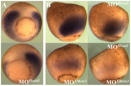
Figure 6. Synergistic effects in loss-of-function of Hoxd1 and XMeis3.
(A) Embryos were injected with 362 ng of MOHoxd1 into the lateral marginal zone on the left side of embryos, rendering the un-injected side an internal control. Embryos were allowed to develop until control stage 11 and assayed by in situ hybridisation for expression of Hoxd1. Embryos are shown in vegetal view, with dorsal up. Expression of Hoxd1 is reduced on the left side of injected embryos (shown on the bottom of the panel). (B) To investigate whether there is synergy between Hoxd1 and XMeis3, 16 ng MOXMeis3 and 16 ng MOHoxd1 were injected, together and separately, into the animal hemisphere of one-cell stage embryos. The embryos were harvested at st 11 and assayed for expression of Hoxd1 by in situ hybridisation. Embryos are shown in lateral view, with dorsal to the left. Injection of either MOHoxd1 or MOXMeis3 separately leads to a reduction in the early mesodermal expression of Hoxd1. Their co-injection leads to a further reduction in early mesodermal Hoxd1 expression as compared to injection of either MOXMeis3 or MOHoxd1 separately. This suggests that Hoxd1 and XMeis3 work synergistically in mediating establishment of Hoxd1 expression in mesoderm during early gastrula stages.

