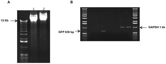
Figure 4. Detection of GFP-coding sequences within genomic DNA prepared from terminally differentiated PC12 cells transduced with RT43.2GFP vectors.
A. Representative gel picture showing genomic DNA fragment with size >10 Kb was used for PCR analysis. Differentiated PC12 cells (5 days with NGF treatment) were exposed to RT43.2GFP vectors for 3 days, and then genomic DNA was isolated and run on a 0.8% agarose gel to purify DNA fragment with size >10 Kb. (lane 2). Non-transduced PC12 cells were used as a negative control as represented by lane 1. B. PCR analysis. The primer pair used for lane 1 (H2O), lane 2 (control PC12 genomic DNA) and lane 3 (differentiated gel purified transduced PC12 genomic DNA) generated a GFP fragment of 636 bp in lane 3. Control primers specific for GAPDH were used for samples in lanes 5–7. Lanes 5 is an H2O control, lane 6 contains DNA purified from control PC12 cells and lane 7 contains gel purified DNA from transduced differentiated PC12 cells; lane 4 is an empty spacer lane.

