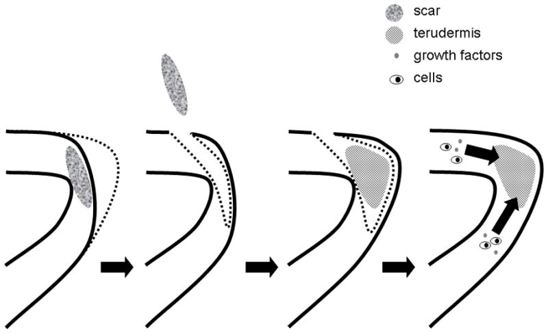
Figure 1.

Schematic illustrating atelocollagen sheet implantation. An incision is made lateral to the scarred site and underlying scar tissue is dissected with a microdissector. Following the removal of scar tissue, the atelocollagen sheet is implanted in a subepithelial pocket. The microflap is put back into its original position and the pocket is closed with a suture or fibrin glue to prevent dislocation of the material. The sheet holds an extended residence time and is infiltrated by cells and possibly growth factors from surrounding tissues. Adapted from [45]
