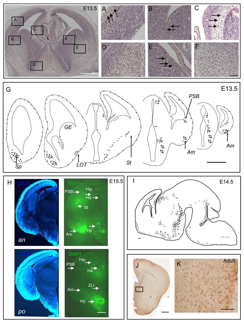
Fig. 2.
Immunohistochemistry for Sizn1 at E13.5, E14.5, E15.5 and adult brains. Top left panel is low magnification image of A–F (Scale bar is 500 µm). (A) Sizn1 expression is found in the molecular layer of the developing cortical plate corresponding to Cajal-Reitzius cells (arrows and see Figure 3). (B) Scattered labeled cells are found at the pallial-subpallial boundary (PSB, arrows). (C) Scattered Sizn1 labeled cells are present in the cortical hem (arrows). Numerous labeled cells are found in the developing septal region (D) and striatum (F). (E) Labeled cells in the medial ganglionic eminence (MGE, arrows) are primarily found in the ventricular zone. Scale bar in image F is 100 µm and corresponded to images A–F. Schematic diagrams of E13.5 brain coronal section (G; Scale bar=500 µm)) and of E14.5 sagittal section (I; Scale bar=100µm) for Sizn1 expression pattern. (H) Coronal section of E15.5 embryonic caudal forebrain. Anterior (an) and posterior (po) DAPI labeled sections on the left and Sizn1 immunofluorescence on the right (green). Sizn1 expression is localized in the amygdala, habenula, hippocampus, hypothalamus, pallium-subpallium boundary and zona limitans intrathalamica (scale bar = 500 µm). (J and K) Immunostaining for Sizn1 in the adult brain with emphasis on the septal region (Scale bar=500 µm (I) and 100 µm (J)). Am=amygdala, Ha=habenula, Hip=hippocampus, Hy=hypothalamus, Sp=Septum, St=striatum, LOT=lateral olfactory tract, GE=gangilonic eminence, PSB=pallium-subpallium boundary and ZLI= zona limitans intrathalamica.

