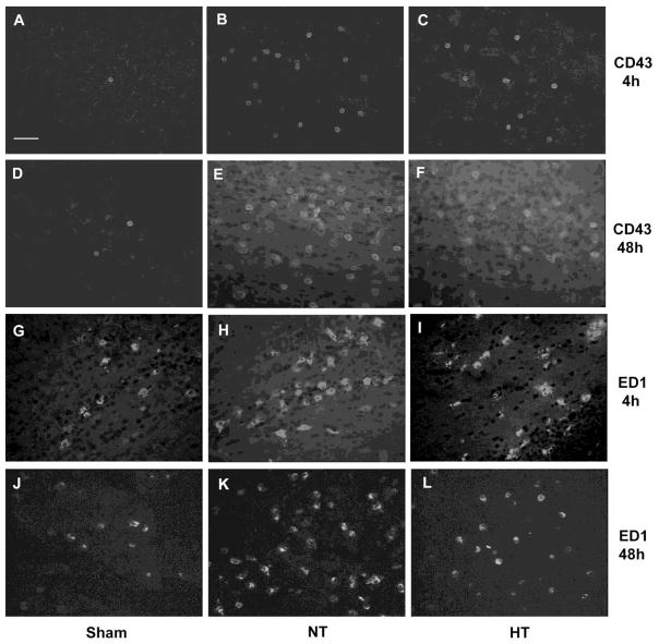
Figur 4.
Immunostaining of CD43 and ED1 in the rat brain at 4 and 48 h after cerebral HI. Immediately after HI, rats were exposed to either a hypothermia (HT) or a normothermia (NT) condition for 4 h. For the 48 h data, the rat pups in the HT group received iN-IGF-1 treatment immediately after the termination of hypothermia. Brain samples were collected after the termination of hypothermia (4 h after HI) or 48 h after HI. Representative images of CD43 immunostaining were taken at the ipsilateral cortical area (A–F), while images of ED1 immunostaining were taken at the ipsilateral cingulum area (G–L). Only a few CD43+ cells (A, D) or ED1+ cells (G, J) were observed in the sham brain at 4 and 48 h after HI. Cerebral HI caused the infiltration of PMN cells (B) and activation of macrophages/microglia (H) at 4 h after cerebral HI. Hypothermic treatment immediately after HI reduced PMN infiltration (C) and macrophage/migroglia activation (I) in the ipsilateral brain. The recruitment of PMN cells (E) and macrophage/microglia (K) was even more evident at 48 h after HI. Hypothermia combined with IGF-1 inhibited the recruitment of PMN cells (F) and macrophage/microglia (L). Scale bar: 50 μm.

