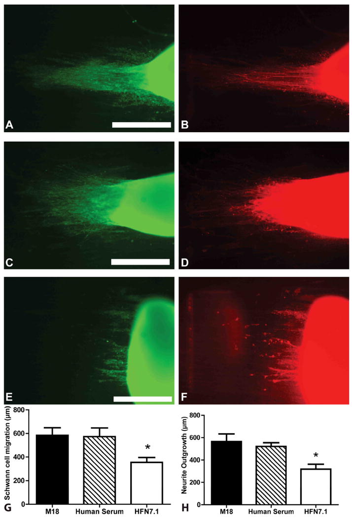
Figure 5.

DRG Schwann cell migration and neurite outgrowth in HFN7.1 antibody inhibited fibers. Migration and outgrowth in M18 isotype control (A, B), normal human serum (C, D), and HFN7.1 antibody incubated fibers (E, F). Left panel is stained for Schwann cell (S100, green) and right panel is stained for neurites (NF160, red) Scale bars = 400 μm. Both controls demonstrated significantly higher Schwann migration and neurite outgrowth when compared to HFN7.1 incubated DRGS (G, H) respectively. *p<0.05. Error bar = Std. dev.
