
Figure 6.
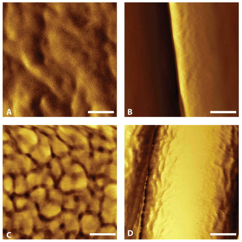
Atomic force microscopy images of PAN-MA polymer topography. (A): Smooth film. (B): Aligned PAN-MA fiber scaffold. (C) Smooth film with fibronectin. (D) Aligned fibers with fibronectin. Scale bar = 100 nm.
Official websites use .gov
A
.gov website belongs to an official
government organization in the United States.
Secure .gov websites use HTTPS
A lock (
) or https:// means you've safely
connected to the .gov website. Share sensitive
information only on official, secure websites.
Atomic force microscopy images of PAN-MA polymer topography. (A): Smooth film. (B): Aligned PAN-MA fiber scaffold. (C) Smooth film with fibronectin. (D) Aligned fibers with fibronectin. Scale bar = 100 nm.