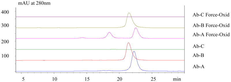
Figure 2.
AEX at pH 7.5 of XOMA 3AB Antibodies. The two more neutral antibodies (Ab-A, pI=7.4 and Ab-B, pI=6.7) bound to the anion-exchange resin while the basic Mab (Ab-C) flowed through. The force-oxidized Ab-A exhibited two more basic peaks. In contrast, force-oxidized Ab-B shows only a single major peak. Both basic peak fractions of Ab-A were subjected to further characterization by peptide mapping (Figure 4). Force-oxidized Ab-B was also characterized by peptide mapping to determine the sites of modification since no apparent resolution of oxidized species was observed.

