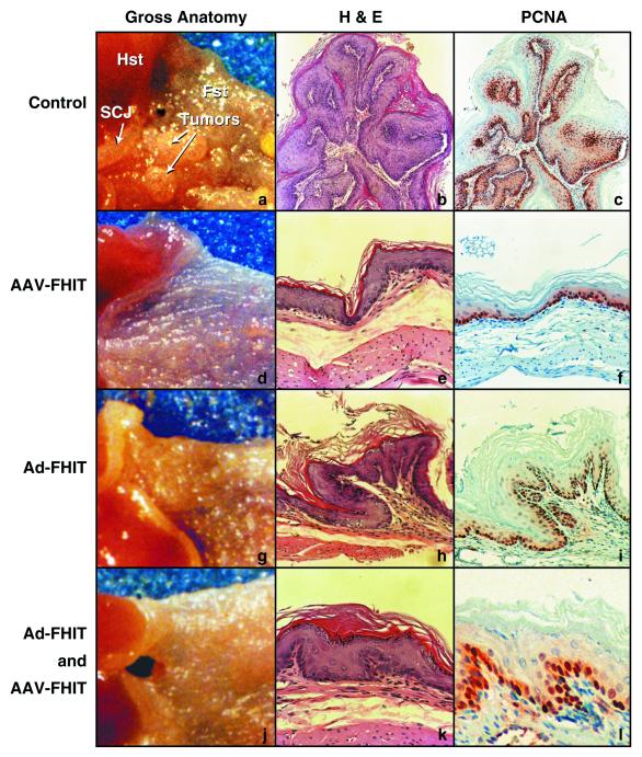
Figure 3.
Gross anatomy and histopathology of murine forestomach after FHIT-gene therapy. Typical aspects of NMBA-induced pathology in the control group (a–c) are compared with the three treatment groups: AAV-FHIT (d–f), Ad-FHIT (g–i), and the combined treatment group Ad-FHIT with AAV-FHIT (j–l). (a, d, g, and j) Gross anatomy of the forestomach and SCJ (magnification ×5). b (magnification ×50) and e, h, and k (magnification ×200) are hematoxylin and eosin (H&E)-stained forestomach sections. c (magnification ×50) and f, i, and l (magnification ×200) depict PCNA immunohistochemistry in forestomach sections adjacent to the corresponding H&E sections. PCNA immunohistochemistry shows abundant, intensely stained cells in S phase in a control forestomach showing a papilloma with hyperplastic epithelium (b and c). In AAV-FHIT (e and f), Ad-FHIT (h and i) and the combined treatment group (k and l), PCNA-positive cells are found mostly in basal cells of the near normal epithelium.

