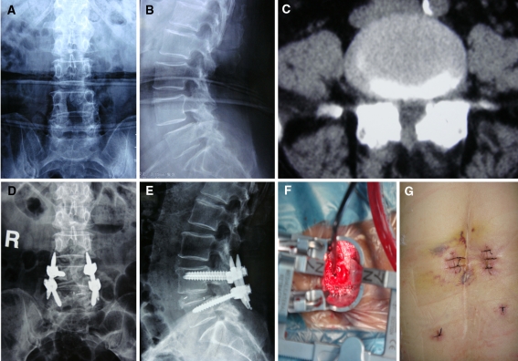
Fig. 1.
Anteroposterior (a) and lateral (b) radiographs of a 71-year-old female with degenerative spondylolisthesis grade 1 after laminectomy. The preoperative CT (c) showed spinal canal stenosis. Anteroposterior (d) and lateral (e) radiographs after MiTLIF and percutaneous pedicle screw fixation showed reduction of the spondylolisthesis. The view under Quadrant system (f) attached with the light cables. The previous large midline skin incision and small skin incision of MiTLIF (g)

