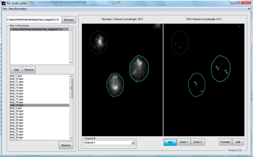
Fig. 1.
FISH Finder editor window showing results from the segmentation process. Segmented nuclei containing two identified FISH signals are outlined in bright green, segmented nuclei containing only one identified signal are outlined in light green and segmented nuclei with no identified signals are outlined in dark green. Signals can be added or removed by selecting the Edit button and then selecting the point of interest on the image using the right mouse click button (signals identified by FISH Finder are labeled by a yellow cross-hair; signals marked by an investigator in FISH Finder's editing screen are labeled by a blue cross-hair). Segmented nuclei can be removed by clicking the middle mouse click anywhere inside the boundary.

