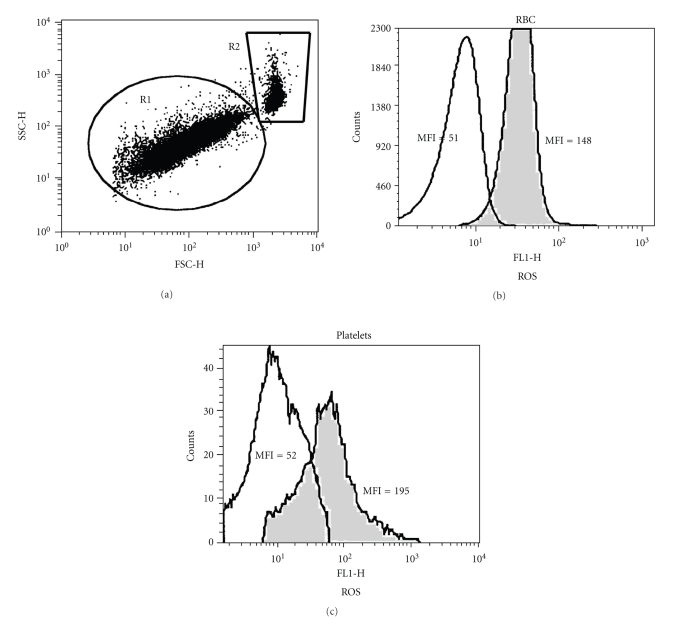
Figure 1.
Flow cytometry analysis of the Epo effect on ROS generation by RBC and platelets. A diluted blood sample obtained from a thalassemia patient was treated for 2 hrs with Epo (1 U/ml) at 37°C, stained with DCF and then stimulated with 1 mM H2O2 for 15 min. (a) FCS versus SSC dot plot. The gates indicate the position of platelets (R1) and RBC (R2). (b-c) Distribution histograms showing DCF-derived fluorescence (FL-1) of untreated (grey) and Epo-treated (white) RBC (b) and platelets (c). The mean fluorescent intensity (MFI) of each population is indicated.

