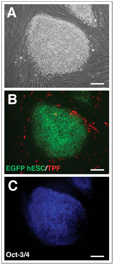
Figure 2.
Oct-3/4 staining of EGFP hESCs grown on TPF. Representative undifferentiated hESC colonies are presented as phase contrast (A); Fluorescent EGFP (hESCs) and Texas Red fluorescence (TPF monolayer) (B); or Oct-3/4 immunostaining (hESCs) (C). EGFP indicates enhanced green fluorescent protein; hESCs, human embryonic stem cells; TPF, term placental fibroblast.

