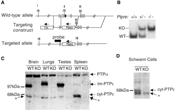
Fig. 1. Generation and characterization of Ptpre–/– mice. (A) Schematic representations of the region of the mouse Ptpre gene chosen for targeting (top), targeting construct (middle) and recombinant allele (bottom). Homologous recombination removed three exons (rectangles), including one (black rectangle) containing C277, the catalytic cysteine of the membrane-proximal catalytic domain. Flanking regions are 1.2 (5′) and 10 kb (3′) in length. RI-EcoRI sites in the targeted region. (B) DNA blot analysis of genomic DNA from Ptpre+/+, Ptpre+/– and Ptpre–/– mice. Genomic DNA was digested with EcoRI and probed with the genomic fragment indicated in (A). Fragments originating in the wild-type (WT; 10.0 kb) and targeted (KO; 11.8 kb) alleles, respectively, are indicated. (C) Protein blot documenting lack of expression of PTPε in Ptpre–/– mice. Protein extracts from the indicated organs of WT or KO mice were analyzed using an anti-PTPε antibody (Elson and Leder, 1995a). Antibody also cross-reacts with PTPα as indicated in the figure. Variations in size of tm-PTPε from brain, lungs and testes are due to tissue-specific glycosylation (Elson and Leder, 1995a). Note the presence in several lanes of PTPε-related bands migrating at ∼68 kDa, (asterisk), slightly faster than cyt-PTPε. (D) Protein blot analysis as in (C), documenting expression levels of cyt-PTPε in primary WT or KO Schwann cells derived from 3- to 5-day-old pups.

An official website of the United States government
Here's how you know
Official websites use .gov
A
.gov website belongs to an official
government organization in the United States.
Secure .gov websites use HTTPS
A lock (
) or https:// means you've safely
connected to the .gov website. Share sensitive
information only on official, secure websites.
