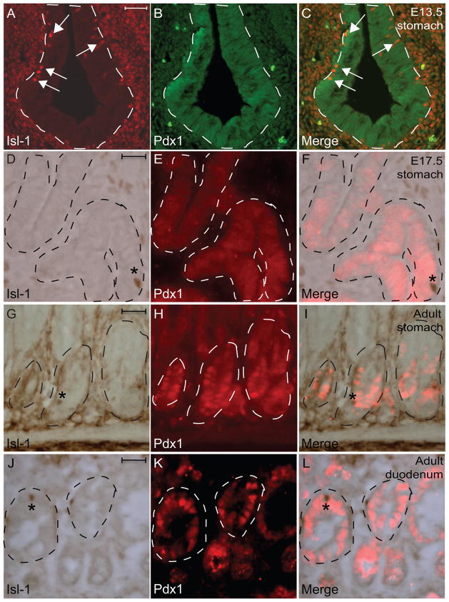
Fig. 6.
Immunohistochemical double-staining for Isl-1 and Pdx1 in the gastrointestinal epithelium. (A–I) Isl-1 with Pdx1 at E13.5 (A–C), E17.5 (D–F) and adult (G–L). Experiments were conducted on the same section. Dashed lines outline epithelial compartments. Arrows in A and C indicate Isl-1+ cells. * in D–L indicates Isl-1+/Pdx1− cells. Scale bars: 25μm.

