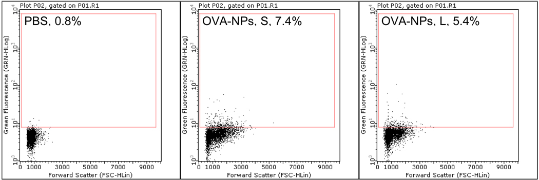
Figure 6.
More draining lymph node cells became fluorescein positive after subcutaneous injection of fluorescein-labeled small OVA-NPs than fluorescein-labeled large OVA-NPs. Fluorescein-labeled small or large OVA-NPs were injected into one hind leg footpad of mice. Popliteal lymph nodes were collected 24 h later, and single cell suspension was analyzed to determine the percentage of fluorescein+ cells (number shown in gated area). Experiment was repeated once with similar result.

