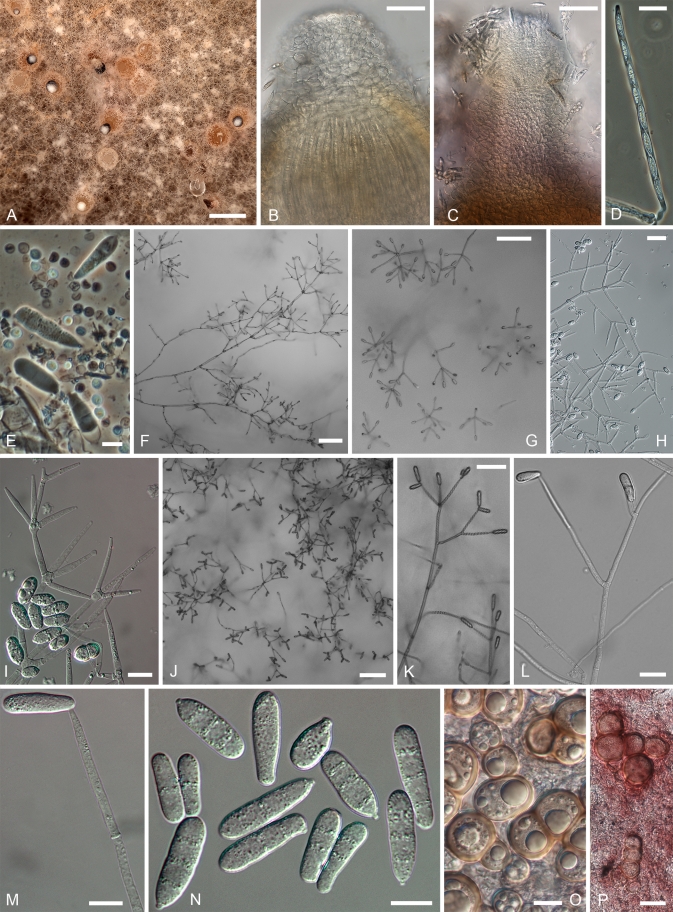
Fig. 18.
Hypomyces gabonensis. A–D. Teleomorph formed in culture. A. Perithecia embedded in the subiculum. B–C. Upper parts of perithecia. D. Ascus. E. Conidia and basidiospores of the host on natural substratum. F–I. Conidiophores with verticillately placed conidiogenous cells and conidia formed first in culture. J–N. Conidiophores with sparingly placed conidiogenous branches and larger conidia produced after the first formed anamorph structures in culture. O, P. Chlamydospores among subiculum. (E. Holotype, TU 112024 on host; A–D, F–P. Ex-type culture, TFC 201156 on MEA; F–N. 12 d; A–D, O. 3 mo; P. 6 mo). Scale bars: A = 500 μm; F, G, J = 100 μm; B, C = 50 μm; H = 25 μm; D, K, L, P = 20 μm; E, I, M–O = 10 μm.

