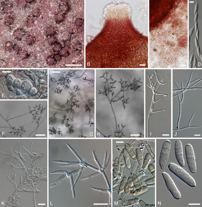
Fig. 8.
Hypomyces virescens. A–D. Teleomorph from a dried culture on MEA. E–N. Anamorph on MEA. A. Perithecia embedded in the subiculum. B. Upper part of a perithecium. C. Base of a perithecium and subicular hyphae. F. Asci and ascospores. E. Chlamydospores among subiculum. F–J. Conidiophores with conidiogenous cells and conidia. K, L. Upper parts of conidiophores. M, N. Conidia. (A–E. Isotype, TU 112905; F–I, K–M. G.A. i1906; J, N INIFAT C10/110). Scale bars: A = 500 μm; F, G = 100 μm; H = 50 μm; B, C, I–L = 20 μm; D, E, M, N = 10 μm.

