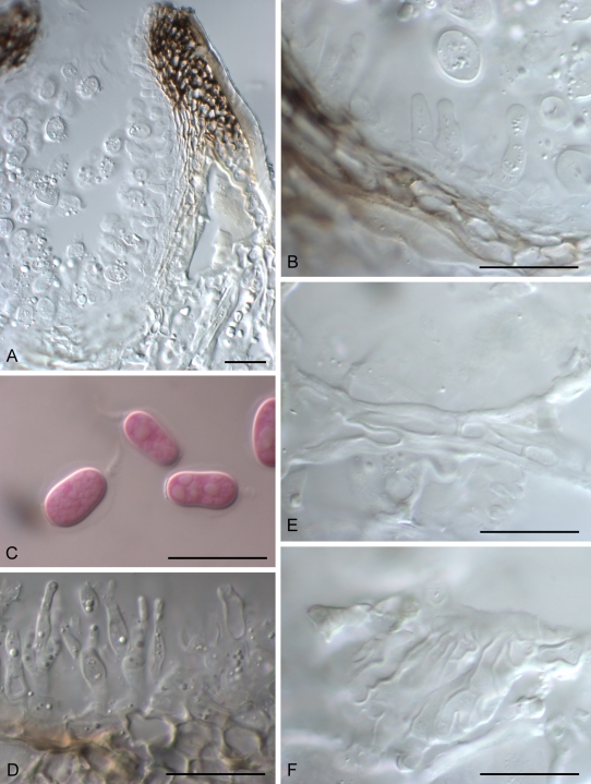
Fig. 4.
Guignardia korthalsellae A. Conidioma in vertical section. B. Wall of conidioma with conidiogenous cells. C. Conidia. D. Conidiogenous cells and conidia of spermatial Leptodothiorella state. E. Fungal hyphae within phylloclade between hypodermal cells. F. Fungal hyphae within phylloclade, showing broad, plate-like layer of hyphae between hypodermal cells. A, B, E, F = PDD 65953; C, D = PDD 94922. Scale bars: A = 50 μm, B–F = 20 μm.

