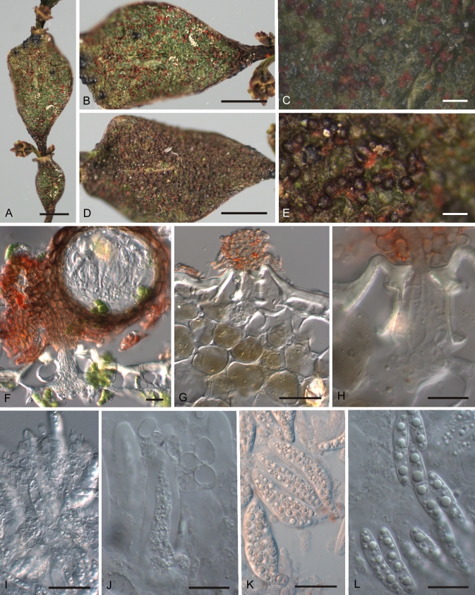
Fig. 6.
Rosenscheldiella korthalsellae A. Infected internodes. B. Detail showing immature, reddish ascomata. C. Detail of B. D. Infected internode densely covered with mature, blackish ascomata. E. Detail of D. F. Ascoma in vertical section, pseudothecium on pad of stromatic tissue developing above stoma. G. Pad of stromatic tissue above stoma, fungal hyphae packing substomatal cavity but otherwise sparse within the internode. H. Detail of G. I. Hymenium, squash mount showing loose, more or less globose cells of hamathecial tissue. J. Detail of hamathecial cells. K. Asci. L. Ascospores. PDD 94885. Scale bars: A, B, D = 2 mm; C, E = 0.5 mm; F = 100 μm; G, I, K = 20 μm; H, J, L = 10 μm.

