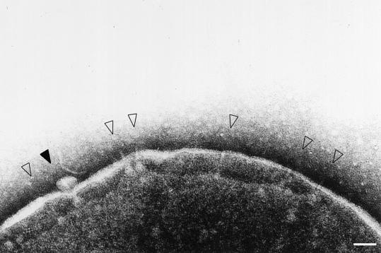
Fig. 1. Electron micrograph of osmotically shocked M94 cells. Cells were negatively stained with 2% phosphotungstic acid pH 7.0 and observed under TEM. The open arrowheads indicate the type III secretion complexes on the bacterial envelope and the closed arrowhead denotes the bleb-like moiety associated with the tip of the complex, reminiscent of the secreted proteins through the type III secretion complexes. Scale bar, 100 nm.

An official website of the United States government
Here's how you know
Official websites use .gov
A
.gov website belongs to an official
government organization in the United States.
Secure .gov websites use HTTPS
A lock (
) or https:// means you've safely
connected to the .gov website. Share sensitive
information only on official, secure websites.
