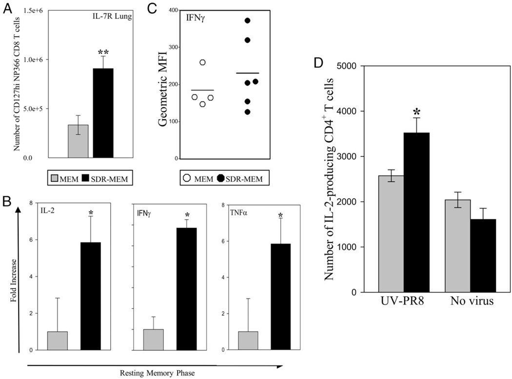
FIGURE 5.
SDR altered cellularity and function of lung parenchymal cells during resting memory. Mouse lungs were sampled 2–3 mo following resolution of a primary A/PR/8 infection. A, In SDR mice, a significant increase in the lung IL-7RHI NP366CD8+ memory phenotype population (**p < 0.05) was noted in the absence of active infection. B, Real-time PCR indicated increases in IL-2, IFN-γ, and TNF-α mRNA expression in lungs at this time. *p < 0.05. C, When lung CD8+ T cells were stimulated ex vivo with NP366–74 peptide and stained for intracellular IFN-γ production, there was a suggestive, but nonsignificant, increase in the intensity of IFN-γ –producing NP366–74 cells isolated from SDR-MEM lungs when mean fluorescence intensity (MFI) values were compared. Line indicates the mean MFI value for each group. D, In an effort to pinpoint the enhanced IL-2–producing lung cell population in SDR-MEM mice seen in B, whole lung preparations were cocultured with UV-PR8 for 6 h and then stained for surface markers and intracellular IL-2. In SDR-MEM mice, there was a significant increase (*p < 0.05) in the number of IL-2–producing CD4+ T cells in lung tissue compared with MEM cell preparations.

