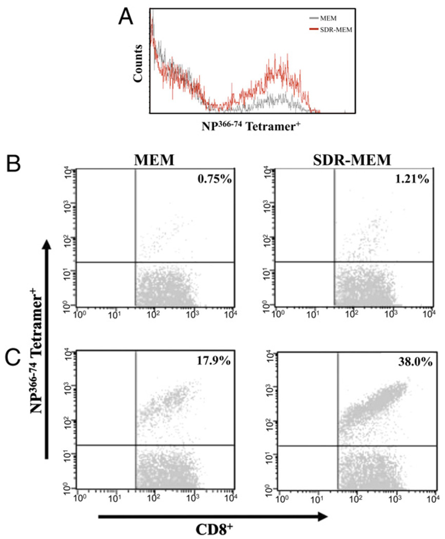
FIGURE 6.
SDR increased the proportion of NP366–74 CD8+ T cells of total CD8+ T cells but did not alter the surface expression of the NP366–74TCR. A, Staining intensity of the NP366 tetramer on memory CD8+ T cells was not changed by exposure to SDR. The proportion of NP366–74CD8+ T cells detected during resting memory (B) and at the peak of the T cell response at day 5 of influenza reinfection (C) was affected by prior SDR experience. Populations shown were sequentially gated on lymphocytes, then CD3+CD8+ T cells, and finally on the NP366–74+ population. Percentages in the upper right quadrant represent the proportion of NP366–74CD8+ T cells to the total CD8+ T cell population.

