Fig. 1.
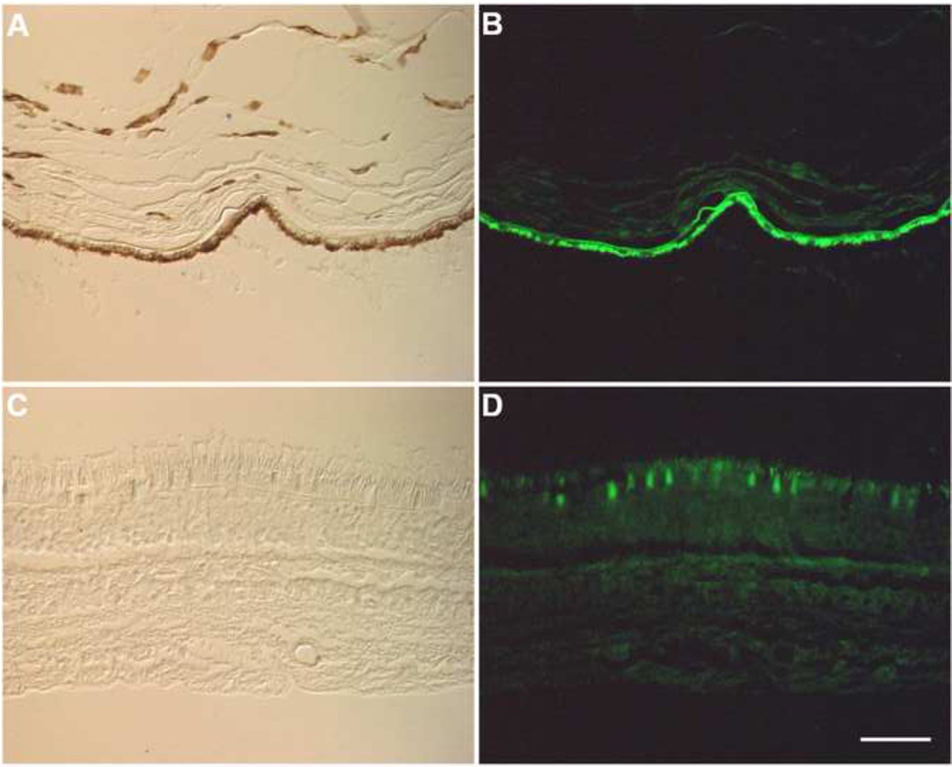
Micrographs of unstained human retina. The sample separated at the RPE-photoreceptor interface during preparation. In panels A and C differential interference contrast images are shown. Autofluorescence images are shown in panels B and D. Panels A and B show the RPE and choroid (note the pigment in panel A) whereas C and D show the neural retina. Although all retinal layers exhibit autofluorescence, RPE cells display bright autofluorescence under conditions used for conventional immunofluorescence using FITC labels (excitation filter, 475nm; emission filter, 530nm). RPE, retinal pigment epithelium; PR, photoreceptor layer; ONL, outer nuclear layer; OPL, outer plexiform layer; INL, inner nuclear layer; IPL, inner plexiform layer; GCL, ganglion cell layer. (Bar=50 µm)

